|
Biological Dosimetry
In the Laboratory of Biological Dosimetry we determine the absorbed dose of ionizing radiation in the human body using various biological dosimetry methods: analysis of dicentric chromosome frequency, the micronucleus assay (manual and semi-automatic), analysis of translocation using the FISH technique, the gamma-H2AX assay and analysis of additional acentric fragments frequency using the premature chromatin condensation assay (PCC). Additionally, we conduct genotoxicity testing in accordance with OECD standards 473 and 487 using chromosomal aberration or micronucleus analysis on cell lines.
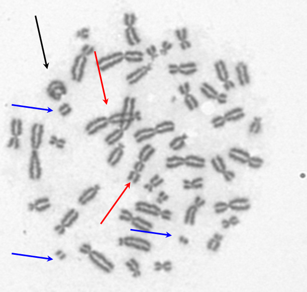
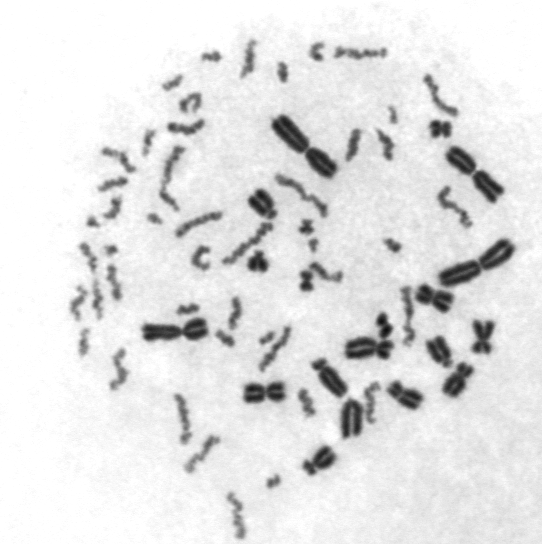
Analysis of Dicentric Chromosome Frequency
Analysis of dicentric chromosome frequency in peripheral blood lymphocytes allows us to assess the absorbed radiation dose of a given individual. Lymphocytes in a collected blood sample are stimulated to divide, cultured for 24 hours, and then fixed and placed on microscope slides. The frequency of dicentrics in unirradiated control blood samples is in the range of 1 dicentric per 1000–2000 cells in the first metaphase after division. Dicentrics are aberrations induced specifically by ionizing radiation and by certain cytostatic drugs with radiation-like effects (radiomimetics). The absorbed dose to the whole body (received over a short period of time – acute dose) is estimated using a calibration curve for cobalt-60 or X-rays (200 kV, 4.5 mA at 200 keV), and a 95% confidence interval is determined. Using standard statistical methods, it is possible to convert a short-term dose into a chronic dose, if it was received over several dozen hours, and to estimate whether the dose was administered to the whole body or only to a portion of it. All calculations are based on the methods included in the ISO 19238:2014 standard.
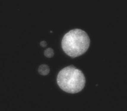
Micronucleus assay
Manual and semi-automatic micronucleus analysis allows for the rapid and precise estimation of absorbed doses in the range of 0.3–3 Gy. Radiation-induced micronuclei are fragments of chromosomes that were not incorporated into cell nuclei during cell division and are visible as morphologically nucleus-like structures in the cytoplasm. The number of micronuclei is proportional to the radiation dose. The procedure for micronucleus assay is similar to the dicentric chromosome assay – a small sample of venous blood is taken. Lymphocytes are stimulated to divide, cytochalasin b is added to block cytokinesis, and the blood is cultured for additional 72 hours. The cells are then fixed, applied to microscope slides, stained, and analyzed manually or semi-automatically (a method 5 times faster but yielding comparable results). The dose is read from a calibration curve for cobalt-60 or X-rays (200 kV, 4.5 mA at 200 keV), and a 95% confidence interval is determined.
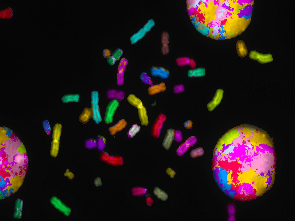
Analysis of translocation
Analysis of translocation using the FISH technique (Fluorescence in situ hybridization) allows for dose estimation. This method is more expensive than dicentric analysis and is primarily used in retrospective dosimetry, when a significant time has passed between irradiation and analysis, such as several months or years. In such situations, the frequency of dicentric chromosomes decreases, while the frequency of translocations remains relatively constant. Analysis of translocation is performed on microscopic slides obtained similarly to the dicentric chromosome test. Chromosomes fixed on microscope slides are hybridized with fluorescently labeled DNA probes, enabling translocation analysis. In our laboratory, we stain three pairs of chromosomes: chromosomes 1, 2, and 3.
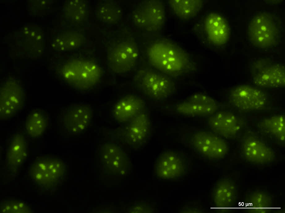
Analysis of the Frequency of Gamma-H2AX Foci
Analysis of the frequency of gamma-H2AX foci allows for the estimation of radiation dose in the range of 0.1–5.0 Gy. The method involves immunohistochemical staining of foci of the phosphorylated version of histone H2AX in human lymphocyte nuclei fixed on microscope slides. Phosphorylation of histone H2AX occurs after DNA damage and is associated with the repair of double-stranded DNA breaks, which are characteristic of ionizing radiation.
Analysis of the frequency of additional acentric fragments (PCC)
Premature Chromosome Condensation (PCC) is a specialized cytogenetic biodosometry technique used to analyse chromosome damage in interphase cells, particularly valuable for rapid assessment and high-dose radiation exposure (> 5 Gy), with results often available in 5–10 hours. Unlike conventional metaphase assays that require 48–72 hours of cell culture, PCC forces interphase chromatin to condense into visible chromosomes, allowing immediate scoring of aberrations, especially in cases where lymphocytes cannot divide due to high-dose radiation.
|
 |